
Lesson 10 – Electron Microscope
Introduction
In this lesson, we will delve into the captivating realm of the electron microscope – a powerful scientific tool that unveils the intricate details of the microcosmos. By harnessing beams of electrons, this device provides unprecedented resolution and magnification, surpassing the limits of traditional light microscopes. Here are the underlying principles, applications, and significance of electron microscopy.
I. Definition
An electron microscope is a type of microscope that utilizes a beam of accelerated electrons for illumination. It is specifically designed to provide high-resolution images and can magnify objects on a nanometer scale. These images are formed through the controlled use of electrons in a vacuum, which is then captured on a glowing screen.
Ernst Ruska (1906-1988), a German engineer and academic professor, constructed the first Electron Microscope in 1931, and the fundamental principles established by his prototype continue to govern modern electron microscopes today.
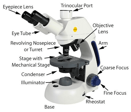
II. Primary parts of an electron microscope
An electron microscope is constructed in the form of a tall, vertically mounted vacuum column and consists of the following components:
1. Electron gun
The electron gun consists of a heated tungsten filament that generates electrons.
2. Electromagnetic lenses
The condenser lens is responsible for focusing the electron beam onto the specimen. Another condenser lens shapes the electrons into a thin and tightly focused beam.
Once the electron beam passes through the specimen, it encounters the objective lens, a set of magnetic coils with high power, which forms an intermediate magnified image.
The final set of magnetic lenses, the projector (ocular) lenses, produces the further magnified and final image. Each of these lenses acts as an image magnifier while maintaining exceptional detail and resolution.
3. Specimen holder
The specimen holder consists of an extremely thin film of carbon or collodion, which is held in place by a metal grid.
4. Image viewing and recording system
The ultimate image is projected onto a fluorescent screen. Below the fluorescent screen, a camera is present to record the image.
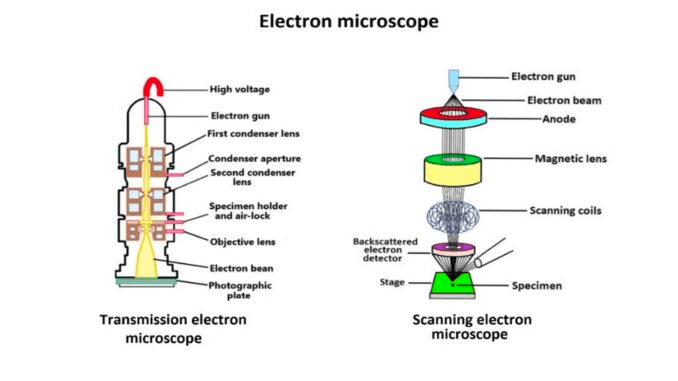
III. Types of electron microscope
1. Transmission Electron Microscope (TEM)
The transmission electron microscope (TEM) is a tool utilized for observing thin specimens that allow electrons to pass through, generating projection images.
The TEM bears many similarities to the conventional (compound) light microscope.
Among its various applications, TEM is commonly used to:
- Visualize the internal structure of cells (in thin sections)
- Examine the organization of protein molecules (enhanced through metal shadowing)
- Analyze the arrangement of molecules in viruses and cytoskeletal filaments (utilizing the negative staining technique)
- Investigate the configuration of protein molecules in cell membranes (via freeze-fracture).
2. Scanning Electron Microscope (SEM)
Conventional scanning electron microscopy relies on the emission of secondary electrons from the surface of a specimen.
With its remarkable depth of focus, a scanning electron microscope functions as the electron microscopy equivalent of a stereo light microscope.
It enables the capture of highly detailed images of cell surfaces and entire organisms, surpassing the capabilities of transmission electron microscopy (TEM). Additionally, it can be utilized for tasks such as particle counting, size determination, and process control.
The term “scanning electron microscope” derives from the image being formed by scanning a focused electron beam across the specimen’s surface in a raster pattern.
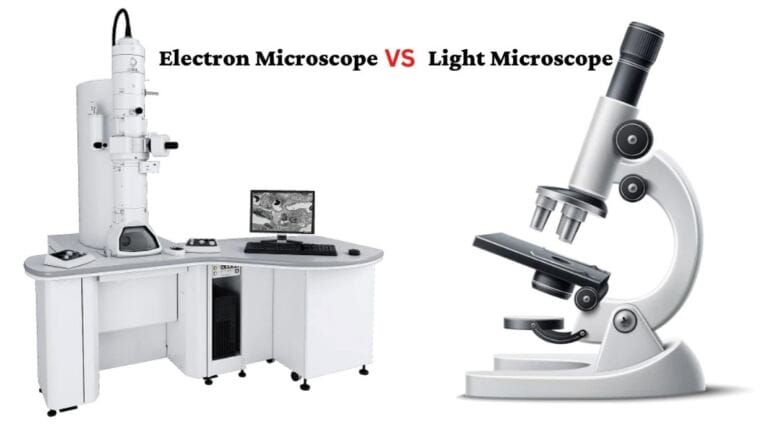
IV. Difference between a light microscope and an electron microscope
1. What is a light microscope?
Microscopes can operate using various methods. The optical microscope, the light microscope, is the most prevalent type. It functions by reflecting visible light onto the sample under observation. Optical microscopes are commonly found in physics or biology labs within educational institutions, such as schools and universities. They serve as valuable tools for demonstrating microscopes’ functionality, operation, and applications.
2. Key differences between light microscope and electron microscope
An electron microscope operates by utilizing accelerated beams of electrons. When these beams interact with the sample, they generate magnetic fields. An optical system, similar to the lenses in a light microscope, employs these magnetic fields to create an image of the observed sample or a specific part of it.
Consequently, electron microscopes offer significantly higher resolution compared to light microscopes. By utilizing electron beams with wavelengths up to 100,000 times shorter than photons, electron microscopes can achieve up to 10,000,000 times better-resolving power than their light microscopy counterparts.
Electron Microscope
- Utilizes beams of electrons to generate an image of the sample.
- Represents a modern advancement over older microscope designs.
- Images are produced through the generation of magnetic fields.
- Provides superior resolution capabilities.
Light Microscope
- Operates by using beams of visible light that illuminate the sample.
- Represents the earliest design of microscopes invented.
- Generates virtual images formed at a greater optical distance than the actual sample-to-lens distance.
- Resolution is constrained by the wavelength of visible light.
V. Uses of electron microscope
Electron microscopes are instrumental in exploring the intricate structure of a wide range of biological and inorganic specimens, including microorganisms, cells, large molecules, biopsy samples, metals, and crystals.
- In industrial applications, electron microscopes are frequently used for quality control and failure analysis.
- Modern electron microscopes employ specialized digital cameras and frame grabbers to capture electron micrographs, resulting in detailed and precise images.
- The field of microbiology owes much of its advancement to the electron microscope. The study of microorganisms such as bacteria, viruses, and other pathogens has significantly contributed to developing highly effective disease treatments.
[Electron Microscope – Definition, Types, Parts, Application, Advantages, Disadvantages.mp4]
Conclusion
In conclusion, the electron microscope has revolutionized our understanding of the microscopic world. Through this lesson material, we have explored the principles, types, and applications of electron microscopy, witnessing its ability to expose the intricate structures and secrets of matter at unprecedented levels of detail. (Paragraph)
Electron microscopes offer significantly higher magnification and resolution, making them essential for studying ultrafine details at the atomic and molecular levels and examining materials and nanoscale structures.